

Thermo Fisher Scientific highlights case for combining electron microscopy and microtomography
In a recent release, Franz Kamutzki, Senior Account Manager at Thermo Fisher Scientific Inc, Waltham, Massachusetts, USA, explored how engineers and researchers can best combine electron microscopy (EM) and micro-computed tomography (microCT) to gain deeper insights into materials at different length scales.
Kamutzki highlighted the technique through the study of functionally gradient materials (FGM), which feature a continuously variable directional composition and structure to enhance performance. These materials are particularly valuable in industrial applications required to withstand extreme stresses (e.g. aerospace components and high-performance coatings). Characterisation of FGMs is necessarily challenging, as their continuous compositional changes require a multi-analytical approach to ensure no variations are overlooked.
Analytical techniques and combined approaches
Each analytical technique provides unique and complementary insights, explains the author. Electron microscopy delivers nanometre-scale resolution, offering detailed information about a material’s surface structure, composition and microstructure. This makes it useful for studying fine structural details that influence material performance.
MicroCT, meanwhile, excels at non-destructive 3D imaging of larger samples, revealing internal features such as porosity, cracks and density variations. While its resolution is lower than EM, it provides a macroscopic perspective that allows researchers to understand how internal structures and defects are distributed throughout a material.
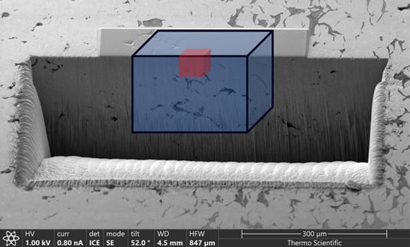
By integrating EM and microCT, engineers and researchers can achieve a multiscale understanding of materials that neither technique can deliver in isolation. Bridging the gap between macro- and nanoscale analyses allows for comprehensive visualisation, with MicroCT’s 3D imaging maps showcasing large-scale defects, with EM adding detailed insights into smaller features such as grain structures and elemental composition.
Combining data from both techniques also enables the accurate identification of defect locations, sizes and types. The insights gained from this hybrid approach can help engineers to refine manufacturing processes, thus improving material properties and performance.
Kamutzki used cold sprayed additively manufactured copper-nickel (NiCu) alloys, used in rocket chambers, to exemplify a use case of the combination technique. As FGMs, using only one method of analysis could lead to engineers missing critical material property details, thus causing safety issues.
In the ‘Multi-scale Characterization of Functionally Gradient Bimetallic Ni-Cu CSAM Alloys’ lecture presented at the 4th Symposium on Materials and Additive Manufacturing, researchers outlined a methodology for combining the two techniques to best achieve a complete picture of materials.
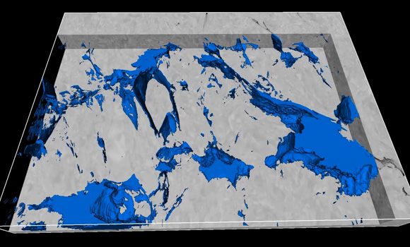
Yorston et al employed MicroCT to visualise porosity distribution in 3D, revealing trends such as increased pore density in nickel-rich regions. Meanwhile, the team’s EM provided nanoscale data on defect morphology and elemental distribution, uncovering smaller defect classes that microCT could not detect. These insights informed adjustments to the alloy composition and processing parameters, ultimately enhancing material reliability.

Adoption barriers
In any industries that use additively manufactured alloys such as aerospace, automotive and energy, a combined microscale analysis approach ensures a thorough understanding of material properties. In these cases, the hybrid approach can help to accelerate iterative development and reduce failure rates.
Despite its potential, however, the widespread adoption of combined EM and microCT analysis faces challenges. For instance, in industrial applications where operators are not necessarily experts in specific microscopy techniques, this approach may be perceived as overly complex. Others may question its scalability for routine industrial use that requires the analysis of thousands of samples
In his paper, Kamutzki noted the suitability of Thermo Fisher Scientific’s Arizo software in overcoming these barriers by adopting a combined data correlation/visualisation approach regardless of scale or modality. Avizo provides digital imaging-based workflows to facilitate materials characterisation and quality control from a single environment. The software offers advanced automation capabilities, allowing the creation of repeatable, reliable workflows to conduct analysis at scale.
The inclusion of artificial intelligence may enable engineers who aren’t confident with image processing to save time on complex analysis while ensuring results consistency. In addition, Thermo Fisher’s dedicated support team, training sessions and how-to guides work to reduce the learning curve for non-expert operators.
By bridging the gap between these two methods, Avizo makes the combined power of microCT and EM both practical and scalable, in an effort to drive faster, more informed decision-making in materials analysis.